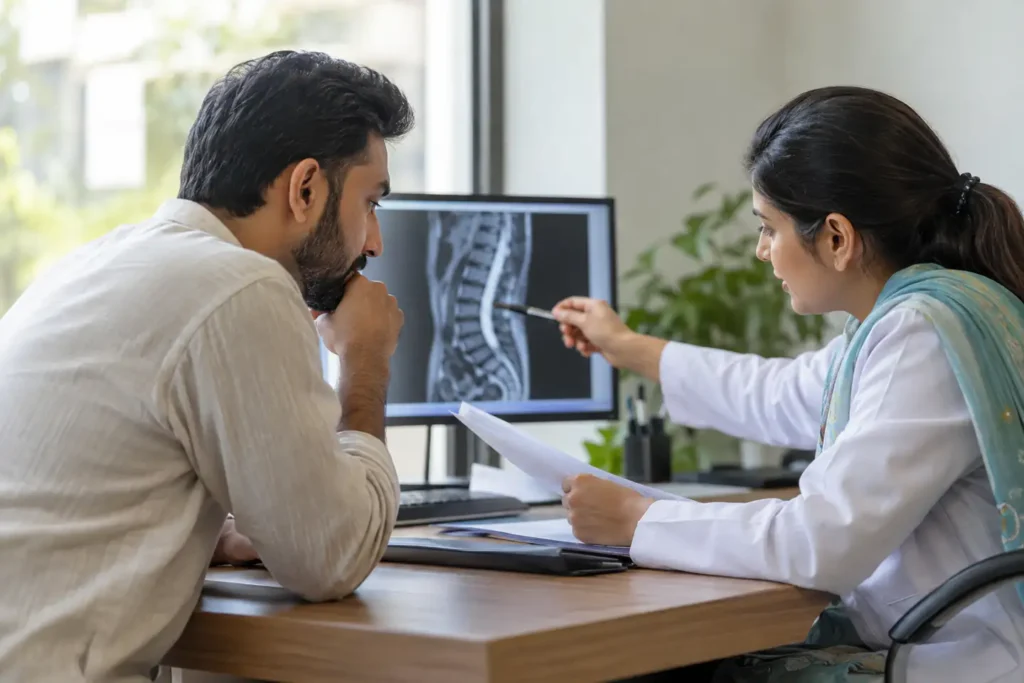
MRI for back pain sounds reassuring when symptoms are severe and you want an exact answer. You may hope a scan will reveal one visible defect and finally explain every ache. But a scan does not measure pain, and many familiar findings—including disc bulges—also appear in people who feel completely well. The more important question is whether imaging would change a clinical decision.
MRI for back pain is generally unnecessary for many uncomplicated episodes without red flags or progressive neurological findings. Imaging may be appropriate when there is suspicion of serious disease, significant neurological impairment, or persistent symptoms for which results would change specialist management. Scan findings must be interpreted alongside clinical symptoms and examination, because common degenerative changes also occur in people without pain.
At a Glance
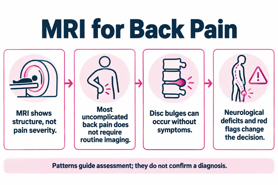
- MRI shows structure, not pain severity.
- Most uncomplicated back pain does not require routine imaging.
- Disc bulges can occur without symptoms.
- Neurological deficits and red flags change the decision.
- Imaging is most useful when it can alter management.
- A clinical examination remains essential.
What Does a Lumbar MRI Actually Show?
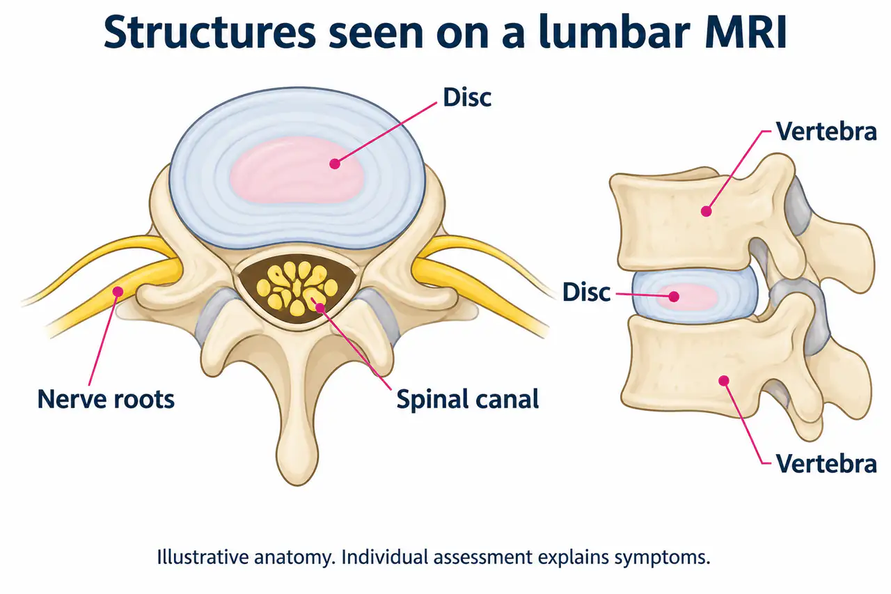
MRI uses magnetic fields to create detailed images of discs, bones, nerve structures and other soft tissues. It can help characterize a suspected disc herniation or certain serious medical conditions. Yet it is a structural test, not a photograph of pain itself. The image does not show exactly which everyday activity you can tolerate or how much recovery is possible.
A report may describe narrowing, dehydration or a disc protrusion even when the patient has only mild symptoms.
Ask what clinical question the scan is meant to answer, not simply whether every word in the report sounds abnormal.
An MRI is a picture of anatomy at a particular time. It can display discs, spinal canal size, vertebrae and nerve structures in much more detail than a plain radiograph, but it cannot directly measure pain or predict how confidently someone can bend or walk. Before asking for one, consider what question you and the clinician are trying to answer. A scan ordered only to identify 'any abnormality' may reveal multiple common changes without clarifying which one is relevant. The value of the result depends on how it changes a real decision.
Why Do Many People Not Need an MRI Immediately?
International guidance advises against routine imaging for uncomplicated low back pain in a non-specialist setting. Many people improve with time, education and appropriate activity, and early imaging can expose incidental findings that increase worry without improving decisions. These recommendations do not mean that imaging is never useful.
Someone has a new movement-related back ache after an unusual lifting day, no nerve deficits and steady improvement over two weeks.
In such cases, an appropriate clinical assessment and monitoring may be more informative than an immediate scan.
For an assessment-led explanation of related symptoms, see WHPT Pakistan’s slipped-disc and disc-herniation rehabilitation resource. It explains what an individualized assessment and rehabilitation pathway may involve.
For recent uncomplicated low-back pain without signs of serious disease, major guidelines advise against routine early imaging. Many episodes improve without a scan, and results showing ordinary degeneration can create unnecessary alarm. Delaying imaging in this context does not mean symptoms are imaginary or that examination is unnecessary. The person still needs a clear history, screening for red flags and a plan to maintain manageable activity. If the presentation changes, the imaging decision can change. The correct message is 'not routinely now,' not 'never.'
What Findings Commonly Appear in Pain-Free People?
Disc bulges, degeneration and other age-related changes become more common over time. A systematic review found many such features in people without back pain. This does not mean MRI findings are always irrelevant, but it does prevent the assumption that a change must be the cause of symptoms solely because it was detected.
An older adult may have several disc bulges on MRI and still walk comfortably, while another person with fewer changes may feel substantial pain.
Interpret the scan’s level, side and severity in relation to symptoms, neurological signs and functional limitation.
Research has found disc bulges and other degenerative findings in many people who have no back pain. That observation does not mean MRI findings are always irrelevant: a focal lesion matching neurological deficits and side-specific leg symptoms can matter enormously. It means the report should be interpreted alongside a physical examination. A phrase such as 'mild posterior bulge' describes an appearance, not a permanent disability. Ask the clinician to show how the finding relates, or does not relate, to the actual symptoms before deciding to stop ordinary movement.
A practical comparison
| What you notice | Why it may matter |
|---|---|
| New uncomplicated back pain, stable examination | Routine early MRI often adds little. |
| Suspected serious pathology or progressive weakness | Medical/specialist evaluation may require urgent imaging. |
| Persistent radicular symptoms and procedure planning | Specialist imaging may guide intervention. |
When Is an MRI More Likely to Change Care?
Imaging becomes more relevant when examination suggests serious disease, significant or progressive nerve compromise, or a specialist is considering an intervention and needs anatomical detail. A prior cancer history, infection risk, trauma or unexplained systemic findings may prompt a different imaging pathway.
Someone develops new progressive leg weakness and altered bladder function: that is not a situation for routine outpatient back exercises.
Urgent assessment determines the type and timing of imaging; a physiotherapist should not delay emergency referral while arranging ordinary exercises.
Imaging becomes more compelling when there is a strong suspicion of cancer, infection, fracture, cauda equina syndrome, or important progressive neurological deficit. Specialist imaging can also help when persistent nerve-root symptoms are being evaluated for procedures or surgery and the result is expected to influence that decision. Different concerns may require different investigations rather than always a routine lumbar MRI. For someone with rapidly developing weakness or bladder dysfunction, emergency assessment matters more than booking a scan privately and waiting to interpret it alone.
For clinical decisions about whether lumbar imaging could change management, see ACR Appropriateness Criteria: Low Back Pain. The decision depends on medical context and whether the scan result would change care.
Does an MRI Confirm Sciatica?
Sciatica is a clinical pattern involving leg symptoms and possible neurological findings. MRI can reveal whether a disc or narrowing might be affecting a relevant nerve root, but an imaging abnormality only strengthens the explanation when the side, level and findings agree. A normal-looking or inconclusive scan does not invalidate a person’s pain.
A right-sided disc finding may not explain left-leg pain, and multiple findings can complicate interpretation.
Use imaging alongside symptom mapping and neurological examination rather than as an independent verdict.
A lumbar MRI may show nerve-root compression, but sciatica is a clinical syndrome, not an image diagnosis. Some individuals have pronounced leg pain with limited visible compression; others have obvious abnormalities with few symptoms. A clinician also assesses strength, sensation, reflexes and provocative maneuvers. If the painful side does not correspond to the finding, the discrepancy should be discussed rather than automatically attributing everything to the scan. Sometimes the examination points toward hip, deep-gluteal or another source requiring a different question.
For examples of why scan findings should be matched with symptoms, see WHPT’s sciatica information when pain follows a nerve-like pattern.
What Should You Ask When You Receive a Report?
Imaging language can be technical and alarming. Ask which findings are expected with age, which fit your symptoms, whether there are findings requiring urgent action, and how the result changes the plan. If management remains the same, another scan may add little. Clinical context is the key to appropriate decisions.
A patient reads “L4-L5 diffuse bulge” and assumes lifting will never be safe again.
Request a plain-language explanation and specific functional guidance instead of treating the report as a restriction on everyday life.
When reviewing a report, identify the spinal level, whether the finding is central or on one side, and whether it describes contact, displacement or compression of a relevant nerve. Ask what the terms 'bulge,' 'protrusion,' 'extrusion' and 'stenosis' mean in plain language. Also ask whether the report fits the neurological examination and whether it changes the proposed next step. Avoid comparing your scan sentence-by-sentence with an online stranger's MRI. Two people with similar radiological words may have very different symptoms, goals and treatment needs.
What Can You Do While Waiting for an Imaging Decision?
If a clinician has screened for urgent problems, comfortable walking and modified daily activity may be reasonable. Avoid staying in bed solely because an MRI has been discussed.
Write down changes in walking, sitting, sleep and leg sensation, and report any new weakness or bladder changes promptly. Those clinical details help decide whether imaging is necessary.
While a clinician is deciding whether imaging is indicated, stable symptoms can often be managed with appropriate advice, comfortable movement and a modified workload. This is not a substitute for urgent medical care when warning signs appear. Keep a concise record of functional changes, including sitting duration, walking distance and any changes in leg strength or sensation. Such information helps show whether a conservative plan is progressing or whether further investigation is necessary. Repeated scans are rarely useful when no clinical question has changed.
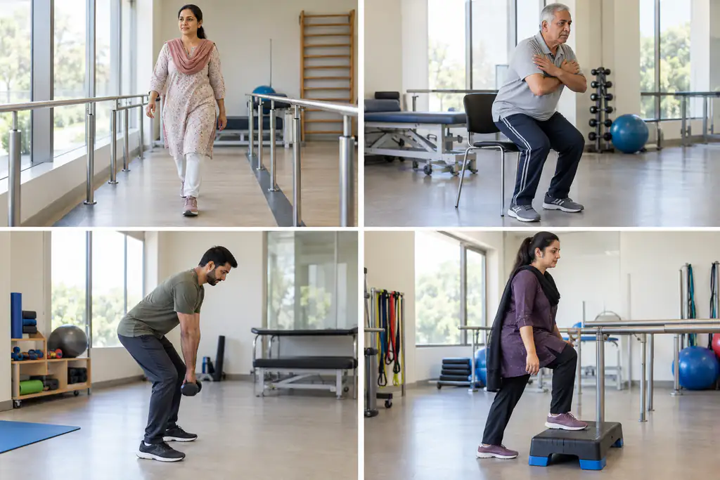
If medical evaluation supports conservative care, therapeutic exercise is planned around what the person can actually do rather than the image report alone.
How Are Scan Findings Combined With Physiotherapy?
The physiotherapist considers how symptoms behave, movement tolerance, strength, sensation and reflexes, plus the reason imaging was ordered. Treatment decisions depend on the entire picture.
Someone with a stable disc finding and improving function may need a graded rehabilitation plan, whereas worsening nerve findings warrant medical review regardless of the original report.
A physiotherapy consultation after imaging should connect the anatomy to function. An L4-L5 report is only one part of the picture; the examination might show that a person's principal difficulty is sustained sitting, lifting or a particular neurological sign. Rehabilitation can then be adjusted for irritability and gradually build tolerance. If a report suggests significant compression consistent with worsening deficits, medical or surgical input may take precedence. The scan should support the decision-making conversation, not replace assessment or be used to frighten someone away from all exercise.
Which Symptoms Need Urgent Medical Imaging Assessment?
Saddle numbness, altered bladder/bowel control, severe or progressive leg weakness, suspected infection, significant trauma or other major red flags require urgent medical evaluation.
Seek urgent medical assessment for new difficulty passing urine, loss of bowel or bladder control, numbness around the groin or saddle area, rapidly worsening or significant weakness in one or both legs, or new symptoms affecting both legs. These can signal serious nerve involvement. Back pain after significant trauma, pain with fever or marked illness, or unexplained symptoms alongside a serious medical history also requires prompt medical attention rather than trial-and-error stretching.
A scan should never be used as reassurance if serious symptoms develop afterward; new clinical findings can change the urgency immediately.
Seek urgent evaluation for new urinary retention, loss of saddle sensation, rapidly worsening or bilateral leg weakness, suspected serious infection, significant trauma or other serious neurological changes. A person with less urgent persistent leg pain that has not improved with appropriate care may need a specialist opinion and selected imaging. When symptoms are stable and improving, a repeated MRI seldom provides a better measure of progress than strength, function and symptom trends. The urgency of imaging depends on consequences of delay, not the intensity of curiosity about the scan.
Frequently Asked Questions
Can an MRI show the exact cause of every back pain episode?
No. MRI shows anatomy and some abnormalities but cannot reliably identify every painful structure or quantify pain intensity.
Does a disc bulge on MRI mean surgery?
No. Many disc findings are managed nonsurgically. Surgery decisions depend on symptoms, neurological findings and specialist assessment.
Is an X-ray better than MRI for back pain?
They answer different questions. X-rays show bones differently; MRI shows discs and neural tissues. Imaging choice depends on the clinical problem.
Should I get another MRI if my back hurts again?
Not automatically. A clinician considers whether symptoms or examination changed enough that another image would alter the care plan.
Does a lumbar MRI expose me to radiation?
No. MRI uses magnetic fields and radio waves rather than ionizing X-rays. It still requires safety screening for some implants, devices or metal fragments. Whether a scan is useful depends on the clinical question and whether its result is likely to change management.
What should I bring to an appointment to discuss MRI results?
Bring the written report, available images, dates of previous scans and a concise symptom timeline. Note any changes in leg strength, sensation, walking, sleep and daily tasks. The clinician should interpret imaging alongside the examination, not in isolation.
What if my scan looks severe but I feel better?
Discuss the finding with your clinician, but improvement in walking, strength and function is meaningful. Radiology terminology does not automatically dictate how restricted a person should be.
The Right Scan at the Right Time
Bring your MRI report to a clinician who can compare its findings with symptoms, strength and actual day-to-day function.
For relevant assessment and rehabilitation options, explore WHPT Pakistan’s back pain physiotherapy. Contact the clinic by WhatsApp or phone if you need help choosing the appropriate appointment.
MRI can be valuable when it answers a real clinical question. When it does not, careful assessment, appropriate activity and monitoring often provide a more useful beginning than an incidental image finding.
The most useful result of a scan is an answer that changes what you do next. Sometimes that answer is the identification of a lesion requiring specialist attention. Sometimes it is reassurance that an image finding is common and does not explain the symptoms. Discuss the result with a clinician who can correlate it with examination and goals, and ask explicitly whether the proposed management would differ if imaging had not been obtained. That question helps patients avoid expensive investigations that add detail without improving decisions.
References
- American College of Radiology. ACR Appropriateness Criteria: Low Back Pain.
- NICE. Low back pain and sciatica in over 16s: assessment and management (NG59).
- Brinjikji W, et al. Spinal degenerative findings in asymptomatic populations. AJNR. 2015.
- MedlinePlus. Herniated disk: patient overview, diagnosis, and management.
- NHS. Back pain: self-care and symptoms requiring medical advice.

About Dr Salman Sabir PT
Dr Salman Sabir PT is a consultant physiotherapist, clinical leader and co-founder of WHPT Pakistan. He holds an MS in Orthopedic Manual Therapy and is undertaking doctoral study in Exercise Physiology. His work focuses on assessment-led rehabilitation, patient education, progressive exercise and long-term recovery.
Need Help Applying This to Your Situation?
General information cannot account for your injury history, strength, movement, medical conditions or personal goals.
BOOK ASSESSMENTCall +92 334 8205557